


Arthritis of the hip is a pathology of degenerative dystrophy characterized by the destruction of hyaline cartilage. The disease progresses gradually, with pain and reduced range of motion. In the early stages of arthritis, without medical intervention, a few years later, the femoral muscles atrophy.
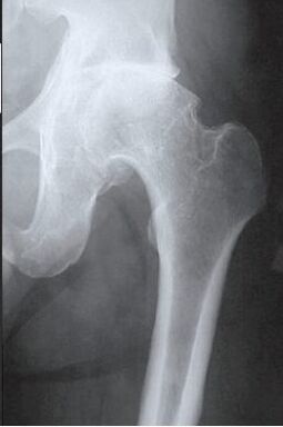 The injured limb is shortened, and the joint space fusion leads to partial or complete hip joint immobilization. The pathological causes are previous injuries, spine curvature, and systemic diseases of the musculoskeletal system.
Osteoarthritis is usually found in middle-aged patients and the elderly. Diagnosis is based on the results of instrumental studies-radiography, MRI, CT, arthroscopy. Pathological treatment of severity 1 and 2 is conservative. If ankylosing scoliosis is detected or medical treatment is ineffective, surgery (arthrodesis, artificial repair) should be performed.
Mechanisms of pathological development
The hip joint is composed of two bones, the lium bone and the femur. The lower part of the body bone is represented by its body. It participates in the joint movement of the femur and forms the upper part of the acetabulum. During exercise, the glenoid fossa is still and the femoral head moves freely. This "hinge" device of the hip joint allows it to bend, straighten, and rotate, and promote hip abduction and adduction. The smooth, elastic, and elastic hyaline cartilage lining the acetabulum and femoral head allows the joint structure to slide unimpeded. Its main function is to redistribute the load during exercise to prevent rapid wear of bone tissue.
Under the influence of external or internal factors, the nutrition of cartilage is disturbed. It does not have its own circulatory system-synovial fluid provides nutrition to the tissues. When suffering from arthritis, it thickens and becomes sticky.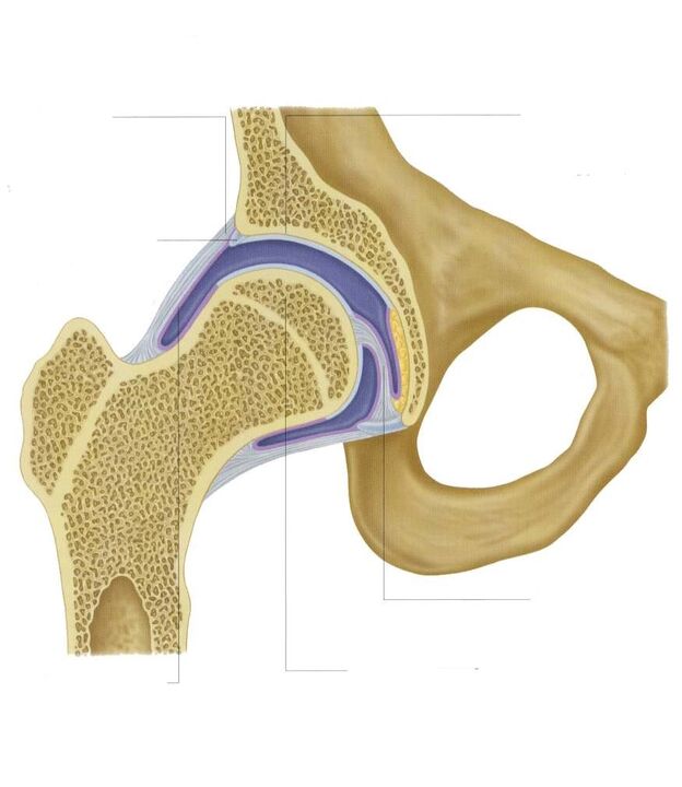 The lack of nutrients causes the surface of the glass cartilage to dry out. It is covered by cracks and causes permanent microtrauma to the tissue during the flexion or extension of the hip joint. The cartilage becomes thin and loses its cushioning properties. The bone deforms to "adapt" to the increase in pressure. In the context of deterioration of metabolism in the organization, destructive and degenerative changes continue to develop.
Causes and provocative factors
Idiopathic or primary arthritis develops for no reason. It is believed that the destruction of cartilage tissue occurs due to the body's natural aging, the slowing of the recovery process, the reduction of collagen and other compounds necessary for the complete regeneration of the hip joint structure. Secondary arthritis occurs in the context of pathological conditions that already exist in the body. The most common causes of secondary diseases include:
- Previous injury-ligament ligament injury, muscle rupture, complete separation of them from the bone root, fracture, dislocation;
- Invasion of joints, the development of abnormal congenital hyperplasia;
- Autoimmune pathology-rheumatoid, reactive, psoriatic arthritis, systemic lupus erythematosus;
- Non-specific inflammatory diseases, such as septic arthritis;
- Special infections-gonorrhea, syphilis, brucellosis, ureaplasmosis, trichomoniasis, tuberculosis, osteomyelitis, encephalitis;
- Destruction of the function of the endocrine system;
- Degenerative dystrophic pathology-osteochondrosis of the femoral skull, osteochondritis dissecans;
- Excessive joint movement, due to the production of "super-extensible" collagen, leads to excessive joint movement and ligament weakness.
Since the cause of the development of arthritis may be thrombosis (bleeding in the hip joint cavity), the predisposing factors include hematopoietic dysfunction. The prerequisites for disease onset are overweight, excessive physical activity, and a sedentary lifestyle. Its development is due to improper organization of sports training, lack of trace elements, food and diet high in fat and water-soluble vitamins. Postoperative arthritis occurs years after surgery, especially if it is accompanied by a large amount of tissue removal. The nutrition of hyaline cartilage is plagued by frequent hypothermia, living in an environment that is unfavorable to the environment, and using toxic substances.
Arthritis of the hip joint cannot be inherited. But in the presence of certain congenital characteristics (metabolic disorders, bone structure), the possibility of its development is greatly increased.
symptom
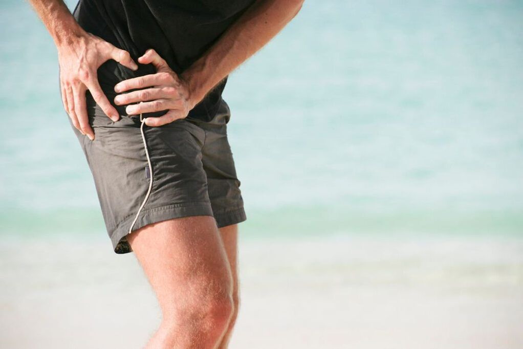
The main symptom of hip joint arthritis is pain when walking in the hip joint area, which radiates to the groin and knee joints. A person's movements are stiff, stiff, especially in the morning. In order to stabilize the joint, the patient starts to walk, and the gait changes. Over time, the limbs are significantly shortened due to muscle atrophy and joint deformation. Another characteristic sign of pathology is restricted hip abduction. For example, difficulties arise when trying to sit on a stool with legs apart.
For the first severe arthritis, there will be periodic pain after intense physical exertion. They are located in the joint area and disappear after a long period of rest.
With the second-degree arthropathy of the hip joint, the severity of the pain syndrome increases. Discomfort can occur even at rest, extending to the thighs and groin, and increasing with weightlifting or sports activity. In order to eliminate the pain of the hip joint, the person hardly starts to walk li. Note the limitation of joint movement, especially when the thigh is abducted and rotated.
Third-degree arthritis is characterized by continuous severe pain, which does not go away during the day and night. Difficulties arise when moving, so when walking, a person is forced to use crutches or crutches. The hip joint is stiff, and the muscles of the buttocks, thighs and legs are obviously atrophy. Due to weakness of the femoral abductor muscle, the pelvic bone is displaced in the frontal plane. To compensate for the shortening of the leg, the patient will lean towards the injured limb when moving. This can cause a strong shift in the center of gravity and increase the stress on the joints. At this stage of the joint, significant joint stiffness develops.
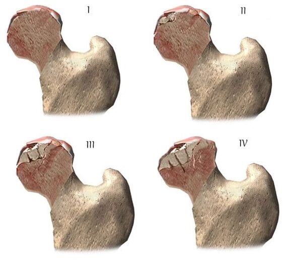
| Bachelor of Science | Radiological signs |
| first of all | The change is not declared. The joint space is moderately narrowed unevenly, and the femoral surface is not damaged. Small bone growth is observed on the outer or inner edge of the acetabulum |
| second | Due to its uneven fusion, the height of the joint space is greatly reduced. The bone of the femur is displaced upward, deformed, enlarged, and its contour becomes uneven. Bone growth on the surface of the inner and outer edges of the glenoids |
| third | The joint space is completely or partially fused. The femoral head is strongly expanded. There are multiple bone growths on all surfaces of the acetabulum |
Diagnostic procedure
When making a diagnosis, the doctor will consider the clinical manifestations of pathology, memory checks, the results of external examinations on the patient, and instrumental research. Radiography is the most useful. With it, the condition of the hip joint, the course of the disease, the degree of damage to the cartilage tissue can be assessed, and in some cases, the cause of its development can be determined. If the phy end of the cervical trunk is enlarged and the acetabulum is inclined and flat, then congenital hyperplasia is likely to occur. Disorders of the shape of the hip bone indicate Perthes disease or epidermal dissolution in adolescents. Imaging examination can reveal the condition of post-traumatic joint disease, even though there is no previous trauma in the memory examination. Other diagnostic methods are also used:
- CT helps to detect the growth of the edge of the bone plate that forms the osteophyte;
- MRI is performed to assess the condition of connective tissue structure and its involvement in the pathological process.
If necessary, check the inner surface of the joint with an arthroscopy. Make a differential diagnosis to rule out gonorrhea, lumbar ac or sternal osteochondrosis. Joint pain can be masked as a clinical manifestation of nerve syndrome caused by nerve entrapment or inflammation. A series of tests can usually be used to rule out neurogenic pathology. Arthritis of the hip joint must be distinguished from trochanteric bursitis, ankylosing spondylitis, and reactive arthritis of the hip joint. In order to rule out autoimmune pathology, biochemical studies of blood and synovial fluid were carried out.
Drug therapy strategy
Medical treatment aims to improve the health of patients. To this end, various clinical and pharmacological groups of drugs are used:
- Non-steroidal anti-inflammatory drugs (NSAIDs)-Nimesulide, Ketoprofen, Diclofenac, Ibuprofen, Meloxicam, Indomethacin, Ketorolac. In order to relieve acute pain, injection solutions can be used, and pills, pills, ointments, and gels can relieve pain of mild or moderate severity.
- Glucocorticoids-triamcinolone, dexamethasone, hydrocortisone. They are used in combination with the anesthetics procaine and lidocaine in the form of intra-articular blockade;
- Muscle relaxants-baclofen, tizanidine. They are included in the treatment plan to treat skeletal muscle spasm and pinch sensitive nerve endings;
- Drugs to improve joint blood circulation-niacin, aminophylline, and pentoxifylline. Prescribe patients to improve tissue nutrition and prevent disease progression;
- Cartilage protector. Only valid in the first and second stages of the joint.
An ointment with a warming effect helps to eliminate mild pain. The active ingredients of the external agent are capsaicin, gall oil, camphor, and menthol. These substances are characterized by local irritation, dispersion, and analgesic effects. Compress the joints with dimethyl sulfoxide, and medical bile can help relieve thigh swelling and swelling in the morning. It is recommended that patients use classical acupressure or vacuum massage to treat arthritis. Daily exercise therapy is an excellent way to prevent the further development of arthritis.
Surgical treatment
Surgery was performed because conservative treatment was ineffective or a pathology complicated with rigidity was diagnosed. Without prosthesis surgery, it is impossible to restore the damaged joint cartilage tissue, but it is necessary to take the correct treatment method, follow all medical prescriptions, maintain a correct lifestyle, perform physical therapy exercises, regular massage courses, take vitamins and appropriateWith nutrition, you can stop the disease process and destroy cartilage and hip joints.