


This material is intended for persons without medical education who would like to learn more about osteochondrosis beyond what is found in popular publications and private practice websites.Patients ask doctors of various specialties questions that are characterized by a complete misunderstanding of the topic of osteochondrosis.Examples of such questions include: "Why does my osteochondrosis hurt?", "Congenital osteochondrosis is discovered, what should I do?" Perhaps this paradigm of illiteracy can be considered a rather common question: "Doctor, I have the first signs of osteochondrosis, how terrible is it?" The purpose of this article is to build material about osteochondrosis, its causes, manifestations, diagnosis, treatment and methods of prevention and to answer the most frequently asked questions.Since all of us without exception are osteochondrosis sufferers, this article is useful for everyone.
What is the structure of an intervertebral disc?
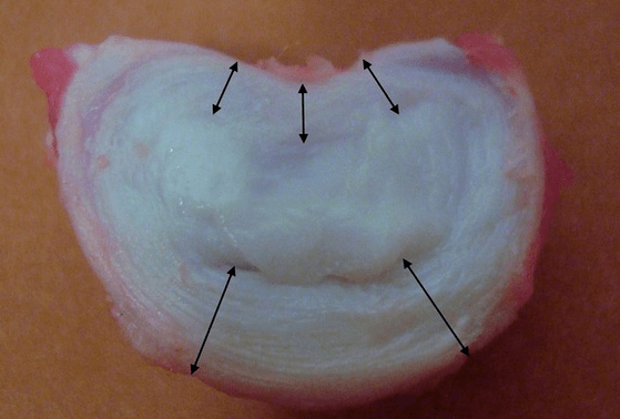
Each intervertebral disc is made up of two distinct parts:
- the outer annulus fibrosus, which is composed of dense fibers that cover the disc from the outside around the periphery;
- The inner elastic component is the nucleus pulposus.
The fibers of the annulus fibrosus are very dense and elastic.As we age, there is a gradual loss of elasticity, and by age 60, the annulus fibrosus becomes stiff.Between the surface of each upper and lower vertebrae and the disc itself lies what is called the endplate, which is the boundary area between the vertebrae and the disc.Because of these endplates, the height of the vertebrae increases, and through them, the nucleus pulposus and disc tissue are extensively nourished by diffusion methods, since the disc cartilage has no blood supply or innervation.
Healthy discs in young people have high metabolic rates.If you introduce contrast into a normal disk, it disappears from it after 20 minutes.
Research shows that the height of each intervertebral disc in adults is approximately:
- 25% of the height of adjacent vertebrae in the neck;
- 20% in the chest;
- 33% are located in the lumbar spine.
That is, in the lumbar region, where the load is greatest, the thickness of the intervertebral disc is greatest.Laboratory studies have shown that a healthy intervertebral disc in young people can withstand static compressive loads of up to 2.5 tons.By the age of 70, this number is reduced to 110 kilograms!That is, the "old and dry disk" is 22 times worse at handling the load to the side and maintaining the increased pressure within the ring.
Why does this happen?Over time, the annulus fibrosus gradually wears away.It can no longer stretch and can only protrude outward, beyond the disc, or break.The core stops transmitting vertical loads and converts them into radial loads.As we age, pressure builds up within the disc and its structure changes.If all of these processes, taking place in a single intervertebral disc, are metastasized throughout the spine, then we have clinically a condition called osteochondrosis.Now we can start defining.
What is osteochondrosis?
It’s scary when the name of this disease is unclear.The medical suffix "-oz" indicates proliferation or enlargement of certain tissues: hyaline degeneration, fibrosis.One example is cirrhosis, when connective tissue grows and functional tissue (liver cells) decreases in size.There may be an accumulation of pathological proteins or amyloid that should not be present under normal circumstances.This storage disease would be called amyloidosis.Due to steatosis, the liver may become significantly enlarged, called fatty liver disease.
Well, it turns out that in intervertebral osteochondrosis, the cartilage tissue of the intervertebral disc increases in volume, because "chondros, χόνδρο" translated from Greek to Russian means "cartilage"?No, rickets, or more accurately, osteochondrosis, is not a storage disease.In this case, the cartilage tissue isn't really growing; we're just talking about changes in the disc structure under the influence of years of physical activity, and we examined above what happens in each individual disc.The term "osteochondrosis" was introduced into the clinical literature in 1933 by A. Hilderbrandt.
Osteochondrosis refers to a dystrophic-degenerative process that is part of the normal aging of the intervertebral discs.None of us would be surprised that a 20-year-old girl's face looks slightly different than her 70-year-old face, but for some reason everyone assumes that the spine and its discs don't undergo the same dramatic temporary changes.Malnutrition is a nutritional disorder, and degeneration is the destruction of the intervertebral disc structure after long-term malnutrition.
Causes of osteochondrosis and its complications
The main cause of uncomplicated physiological osteochondrosis can be considered the person's way of moving: upright walking.Humans are the only species of all mammals on Earth that walk on two legs, and are the only means of locomotion.Osteochondrosis has become a scourge to mankind, but we have liberated our hands and created civilization.Not only did we create the wheel, the alphabet, and master fire thanks to upright walking (and osteochondrosis), but you can read this on your computer screen sitting in the warmth of your home.
Humans' closest relatives, the higher primates - chimpanzees and gorillas - sometimes stand on two legs, but this mode of locomotion is supplementary to them, and most of the time they still walk on four legs.In order for osteochondrosis to go away, as does severe aging of the intervertebral discs, a person needs to change the way they move and eliminate the constant vertical load on the spine.Dolphins, killer whales, and whales do not have osteochondrosis, nor do dogs, cows, and tigers.Their spine is not subject to long-term static and impact vertical loads because it is in a horizontal position.If humans took to the sea, and the natural means of transportation was scuba diving, then osteochondrosis would be defeated.
The upright posture forces the human musculoskeletal system to evolve toward protecting the skull and brain from impact loads.But discs (the elastic pads between vertebrae) aren't the only method of protection.Humans have elastic arches, knee cartilage, and physiological curves of the spine: two lordosis and two kyphosis.All this allows you to not "dump" your brain even while running.
risk factors
But doctors are interested in risk factors that can be modified and avoided to prevent the complications of osteochondrosis, which can cause pain, discomfort, limited mobility and reduced quality of life.Let’s consider these risk factors that are often overlooked by doctors, especially in private medical centers.After all, it is far more beneficial to continually treat a person than to point out the cause of the problem, fix it, and lose the patient.Here they are:
- Longitudinal and transverse flat feet are present.Flat feet cause the arch of the foot to stop bouncing and impact forces are transmitted up the spine without softening.The intervertebral discs are under tremendous pressure and collapse rapidly;
- Overweight and obesity – no comment needed;
- Improper lifting and carrying of heavy objects causes uneven stress on the intervertebral discs.For example, if you carry a bag of potatoes on your shoulders, the powerful load will rest on one edge of the disc and may be too large;
- Lack of physical activity and a sedentary lifestyle.As mentioned above, the greatest pressure on the intervertebral disc is when sitting, because people never sit upright, but always "slightly" curved;
- Chronic injuries, slipping on ice, strenuous lifting, exposure to martial arts, wearing heavy hats, hitting your head on low ceilings, heavy clothing, carrying heavy bags in your hands.
General symptoms
The symptoms described below exist outside of localization.These are common symptoms and can occur anywhere.These are pain, movement impairment, and sensory impairment.There are also phytotrophic diseases or specific symptoms, such as urinary tract disorders, but much less frequently.Let's take a closer look at these signs.
Pain: Muscle and nerve root pain
Pain can be of two types: radicular pain and muscle pain.Radicular pain is associated with compression or herniation or herniation of the disc at the corresponding root at that level.Each nerve root consists of two parts: a sensitive part and a motor part.
Depending on the exact location of the hernia and which part of the root is compressed, sensory or motor impairment may occur.Sometimes two diseases occur at the same time, but to different degrees.Pain is also a sensory disorder because pain is a special sensation.
Radicular pain: Compressive radiculopathy
Radicular pain is familiar to many people.It's called "neuralgia."The swollen nerve root reacts violently to any electrical shock, with pain that is severe and similar to an electric shock.She either shoots in the arms (from the neck) or in the legs (from the lower back).This sharp, painful urge is called low back pain: in the lower back, it's lumbago, and in the neck, it's neck pain, a rarer term.This radicular pain requires forced, analgesic, or analgesic postures.Root pain occurs immediately when coughing, sneezing, crying, laughing, or exerting force.Any impact to the swollen nerve root can cause increased pain.
Muscle pain: myofascial ankylosis
But an intervertebral hernia or disc defect may not compress the nerve root, but can damage nearby ligaments, fascia, and deep back muscles when moved.In this case, the pain will be secondary, sore, permanent, stiff back, and this pain is called myofascial pain.The source of this pain will no longer be nerve tissue, but muscles.Muscles can only respond to any stimulus in one way: to contract.And if the stimulation time is long, the muscle contraction will turn into continuous spasm, which will be very painful.
A classic symptom of this secondary myofascial pain is increased stiffness in the neck, lower back, or thoracic spine, and the development of dense, painful muscle masses—the “rollers” next to the spine, known as the paravertebral spine.In such patients, back pain worsens after working for several hours in the "office", immobile for long periods of time, when the muscles can barely work and are in a state of spasticity.
sensory impairment
If a herniation or hernia occurs, or if a spasmodic muscle presses on a sensitive part of the nerve root, various sensory disturbances may occur.They may accompany the pain or may appear independently after the pain has passed.There are also completely painless sensory disturbances, but these are rare.
Many people are familiar with numbness (reduced sensation or complete numbness) on the tips of the fingers and toes, decreased skin sensitivity, and long streaks that appear like roots.Sometimes there is paresthesia or ant walking, a "goosebumps crawling" feeling.Most commonly, sensitivity disorders occur in the feet and on the tips of the fingers and toes.Sensory impairments are quite unpleasant, but they are not disabling, but movement impairments are likely to be.
peripheral movement disorders
If motor neurons or axons, which are part of the motor part of the nerve, are affected, then muscle weakness or complete immobility can occur.In the second case we are talking about complete paralysis, while in the first we are talking about paresis.Paresis is partial paralysis when muscles cannot function fully.
Most commonly, this disease occurs in the legs, with lumbar herniation and hernias.The motor structures innervate the muscles of the calf and foot.Therefore, in advanced, complex lumbar osteochondrosis, the foot may be spanking.It turns inwards and the person is forced to lift their legs high in order to step with the spanked foot, which is known as the "cock gait".
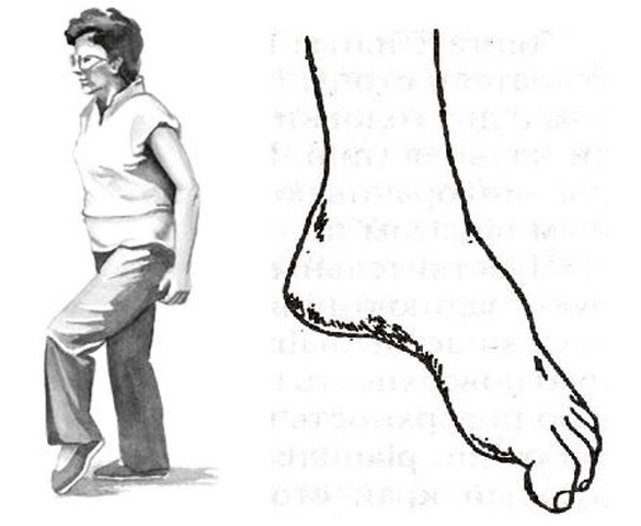
But the whole danger with movement disorders is that they can be isolated and not accompanied by pain.If a person is "pain-free," he or she may not be able to see a doctor in time.Therefore, it is very important for patients with progressive herniation and hernias in the waist to regularly walk on the toes and heels and monitor the work of the muscles.
Local symptoms: main symptoms
Let us now consider the specific symptoms and syndromic features of cervical, thoracic, and lumbar osteochondrosis.We go from top to bottom, from the cervical spine down, through the thoracic spine, to the lumbosacral region.
Diagnosis of osteochondrosis
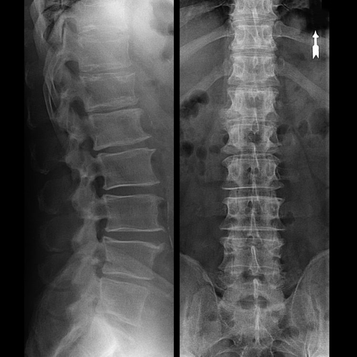
In typical cases, osteochondrosis of the cervical and cervicothoracic spine occurs as described above.Therefore, the main stage of diagnosis was, and still is, identifying the patient's presenting complaint and determining the presence or absence of accompanying muscle spasms by simply palpating the muscles along the spine.Can osteochondrosis be diagnosed through X-ray examination?
An "X-ray" of the cervical spine, even with flexion and extension functional tests, will not show the cartilage because the cartilage tissue transmits the X-rays.Nonetheless, depending on the position of the vertebrae, one can draw general conclusions about the height of the disc, the overall straightening of the physiological curvature of the neck (lordosis), and the presence of growth on the upper edges of the vertebrae and the chronic irritation of its surface by a fragile and dehydrated disc.Functional testing can confirm the diagnosis of cervical spine instability.
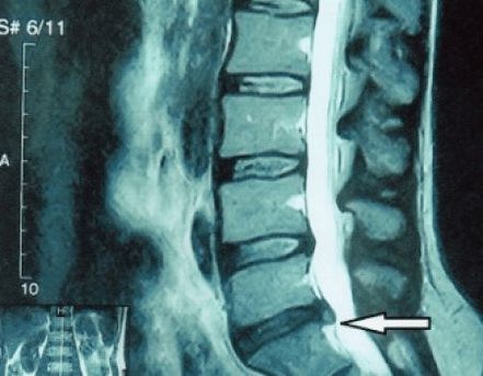
Because the disc itself can only be seen with CT or MRI, magnetic resonance and X-ray computed tomography are used to elucidate the internal structure of the cartilage as well as structures such as protrusions and hernias.Therefore, the diagnosis can be made accurately with the help of these methods, and the tomographic results can be used as an indication or even a local guide for neurosurgical hernia surgical treatment.
Treatment of complications of osteochondrosis
Let us reiterate that there is no cure for osteochondrosis, just like the planned aging and dehydration of the intervertebral discs.You can't make things complicated:
- If you have symptoms of narrowing of the intervertebral disc height, you need to exercise correctly, do not gain weight, and avoid protrusions and muscle pain;
- If there is already a herniation, care needs to be taken not to let it rupture the annulus fibrosus, that is, not to convert the herniation into a hernia, and to avoid the occurrence of multi-level herniations;
- If you have a hernia, then you need to monitor it dynamically, undergo regular MRI examinations, avoid increasing its size, or undergo modern minimally invasive surgical treatment, because without exception, all conservative methods of treating the exacerbation of osteochondrosis leave the hernia in place and only eliminate temporary symptoms: inflammation, pain, shooting and muscle spasms.
But with just the slightest violation of this regime, heavy lifting, hypothermia, injury, weight gain (in the case of the lower back), the symptoms will appear again and again.We will describe how you can deal with unpleasant sensations, pain, and limited mobility in your back in the context of worsening osteochondrosis and an existing herniation or hernia secondary to social ankylosis syndrome.
What should I do when my condition worsens?
Because of acute pain (such as lower back pain), you will need to follow these instructions in the pre-medical period:
- Complete elimination of physical activity;
- Sleep on a firm mattress (orthopedic mattress or firm sofa) to eliminate back sagging;
- It is recommended to wear a semi-rigid corset to prevent sudden movements and "twisting";
- You should place a massage pillow with plastic needles on your lower back, or use a Lyapko massager.It needs to be maintained for 30-40 minutes, 2-3 times a day;
- Afterward, an ointment containing NSAIDs, bee venom, or snake venom can be rubbed into the lower back;
- After wiping, the next day you can wrap your waist with a dry heat wrap, such as a band made of dog hair.
A common mistake is warming up on the first day.This could be a heating pad, a bath routine.Meanwhile, the swelling only increases, and so does the pain.Warmth should only be applied after the "peak of pain" has passed.Thereafter, the heat will enhance the "absorption" of the swelling.This usually occurs within 2-3 days.
The basis of any treatment is symptomatic treatment (eliminating the cause) and etiological treatment (affecting the disease mechanism).It is accompanied by symptomatic treatment.For vertebral pain (caused by spinal problems), here's what happens:
- To reduce muscle and spinal swelling, a salt-free diet and limited fluid intake are recommended.You can even take a mild potassium-sparing diuretic;
- In the acute phase of lumbar osteochondrosis, short-term treatment is possible with intramuscular "injections" of NSAIDs and muscle relaxants: daily.This will help relieve nerve tissue swelling, eliminate inflammation, and normalize muscle tone;
- In the subacute phase, after the maximum pain has been overcome, "injections" should no longer be used, attention should be paid to restorative drugs, such as modern drugs of the "B" group.They can effectively restore damaged sensitivity and reduce numbness and paresthesia.
Physiotherapy measures continue, and the time has come for exercise therapy in osteochondrosis.Its job is to return blood circulation and muscle tone to normal when swelling and inflammation have subsided but muscle spasms have not yet completely resolved.
Exercise therapy (exercise therapy) involves performing therapeutic exercises and swimming.Gymnastics for cervical osteochondrosis do not target the intervertebral disc at all, but the surrounding muscles.Its task is to relieve tonic spasms, improve blood flow, and normalize venous outflow.This is what leads to reduced muscle tone, back pain, and stiffness.
Exercises for the treatment of osteochondrosis must be performed on "warm-up muscles" after a slight general warm-up.The primary therapeutic factor is movement, not the degree of muscle contraction.Therefore, to avoid recurrence, the use of heavy objects is not allowed; gymnastic mats and gymnastic sticks are used.With their help, you can effectively regain your range of motion.
Continue to apply the ointment and use the Kuznetsov applicator.Swimming, underwater massage, and Charcot showers are all on display.In the advanced stages of fading, home magnet therapy and physical therapy medications are required.
Treatment usually takes no more than a week, but in some cases, osteochondrosis can cause dangerous symptoms that may require emergency surgery.
About Shants collar
In the early, acute phase, it is necessary to protect the neck from unnecessary movements.Shant collars are perfect for this.Many people make two mistakes when purchasing this collar.They do not choose it based on their size, which is why it simply fails to perform its function and causes uncomfortable feelings.
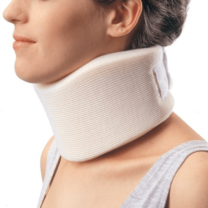
The second common mistake is wearing them for extended periods of time for precautionary purposes.This can lead to weak neck muscles, which will only lead to more problems.For collars, there are only two situations where they can be worn:
- Acute pain and stiffness in the neck that spreads to the head;
- If you are doing physical work while you are healthy, there is a risk of "straining" your neck and aggravating the condition.For example, when you're lying under your car to work on your car, or when you need to reach out and assume an awkward position to wash your windows.
The collar should not be worn for more than 2-3 days, as wearing it for too long can cause venous congestion in the neck muscles, at which point the patient needs to be activated.An analogue of a Shants collar for the lower back is a semi-rigid corset purchased at a plastic surgery salon.
Surgical treatment or conservative treatment?
It is recommended that every patient undergo an MRI after symptom progression, when complications arise, and consult a neurosurgeon.Modern minimally invasive surgeries can safely remove sizable hernias without lengthy hospital stays, without being forced to lie down for days, and without affecting quality of life because they are performed using modern videoendoscopy, radiofrequency, laser technology or cold plasma.You can vaporize some of the kernels and reduce the pressure, thereby reducing the risk of hernias.And you can essentially eliminate the defect, that is, get rid of it completely.
There is no need to be afraid of hernia surgeries; these are no longer the open surgeries of the 1980s and 1990s with their dissection of muscle, blood loss, and subsequent lengthy recovery period.They're more like doing a small puncture under X-ray control and then using modern technology.
Prevention of osteochondrosis and its complications
Osteochondrosis, including complex osteochondrosis, the symptoms and treatments we discussed above, are for the most part not a disease at all, but simply symptoms of the inevitable aging and premature "shrinkage" of the intervertebral discs.Osteochondrosis hardly bothers us:
- Avoid hypothermia, especially in fall and spring, and in winter and fall;
- Do not lift heavy objects, only carry heavy objects in the backpack with your back straight;
- Drink plenty of clean water;
- Don’t get fat, your weight should correspond to your height;
- Treat flat feet (if you have them);
- engage in regular physical exercise;
- Engage in exercise that relieves stress on the back (swimming);
- Quit bad habits;
- Alternate mental stress with physical activity.After every hour and a half of mental work, it is recommended to change the type of activity to physical work;
- You can regularly have at least one lumbar X-ray (two projections) or MRI to see if the hernia (if any) is progressing;
By following these simple tips, you can maintain back health and mobility for a lifetime.